working
with liver cells
in the clinostar
How does the human liver work?
The human liver is a complex and vital organ located in the upper right of the abdomen and is essential for different metabolic processes. It is divided into functional units called lobules, consisting of specialized cells, including hepatocytes, endothelial-, stellate- and Kupffer- -cells in a highly organized 3D structure.
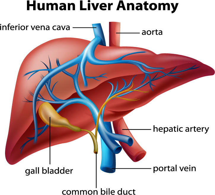
The anatomy of the liver
Blood enters the liver through two large blood vessels, the hepatic artery, and the portal vein. The hepatic artery supplies oxygenated blood to the liver, and the portal vein carries nutrient-rich blood from the digestive system (and this is the only site in the human body where arterial and veinous blood are directly mixed). The blood flows through vessels called sinusoids, which are lined with endothelial cells. Stellate and macrophage-like Kupffer cells have immunological functions and help clean the blood by removing toxins, nucleic acid and peptide fragments, viruses, and even worn-out red blood cells. The blood finally leaves the liver through the hepatic vein, which runs into the inferior vena cava.
How Adelina works with long-term cultures in the ClinoStar
How to work with liver cells in 3D cell culture
Working with liver cells in a 3D environment can provide several advantages over traditional 2D cell culture models. This is because the 3D environment can better mimic the in vivo environment of the liver. This environment brings numerous advantages to the cultures, including improved cell-cell interactions, better cell differentiation, more accurate drug testing, and the possibility of repeated-dose drug toxicity or following liver fibrosis.
One way to use 3D cell culture technology to work with the liver is to create spheroids or organoids. Liver organoids can be generated using various techniques, such as spontaneous or forced aggregation or scaffold encapsulation (in alginate, Matrigel™ collagens, etc.).
3D structures can be used to study many aspects of liver function, including development, disease modeling (including NASH or fibrosis), cancer, drug screening for efficacy, and toxicology.
How to work with liver organoids
in the ClinoStar
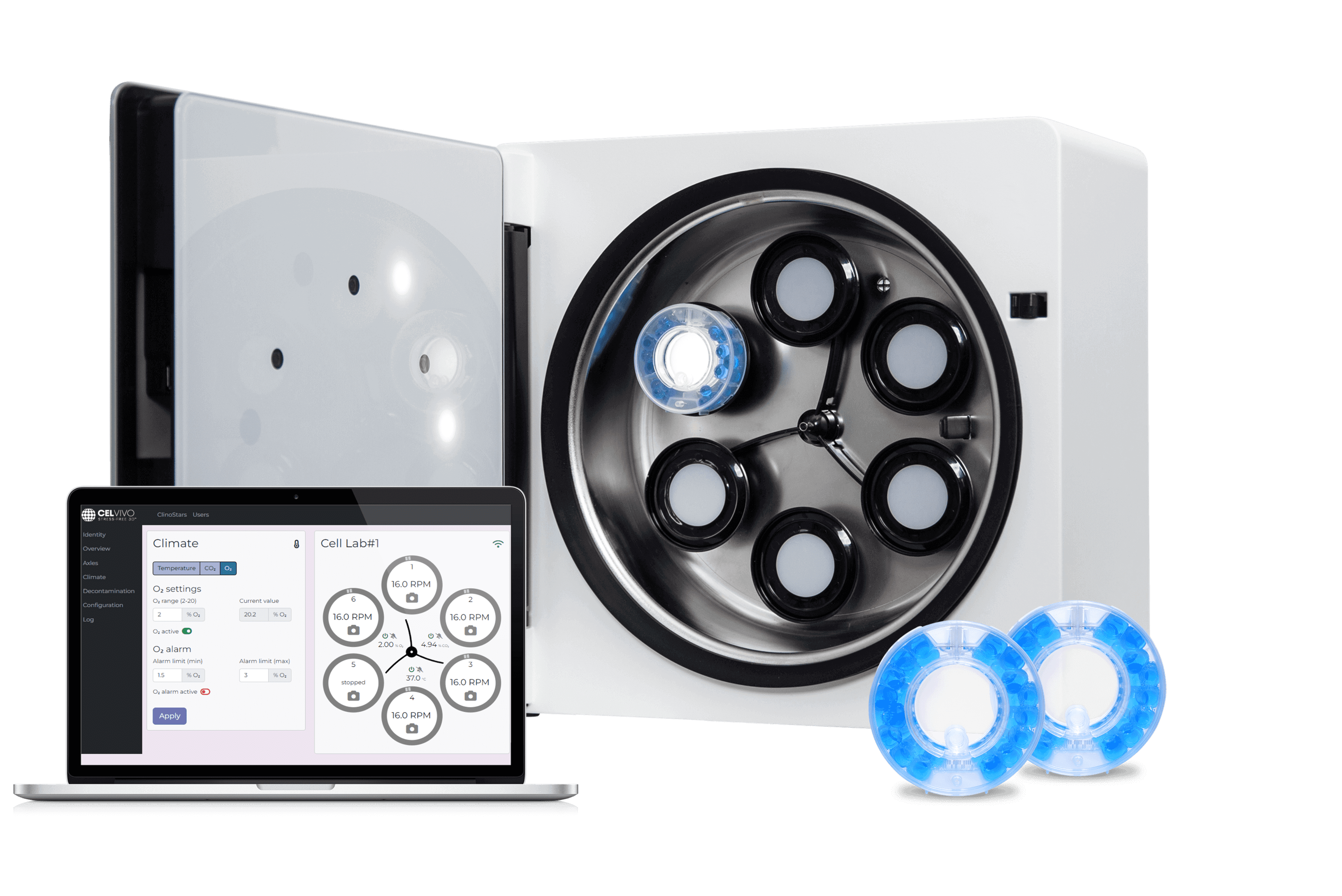
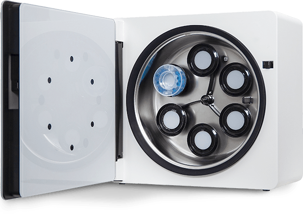
By using the ClinoStar device to study liver cells in a 3D environment, you’ll see how the 3D structure affects the behavior and function of liver cells.
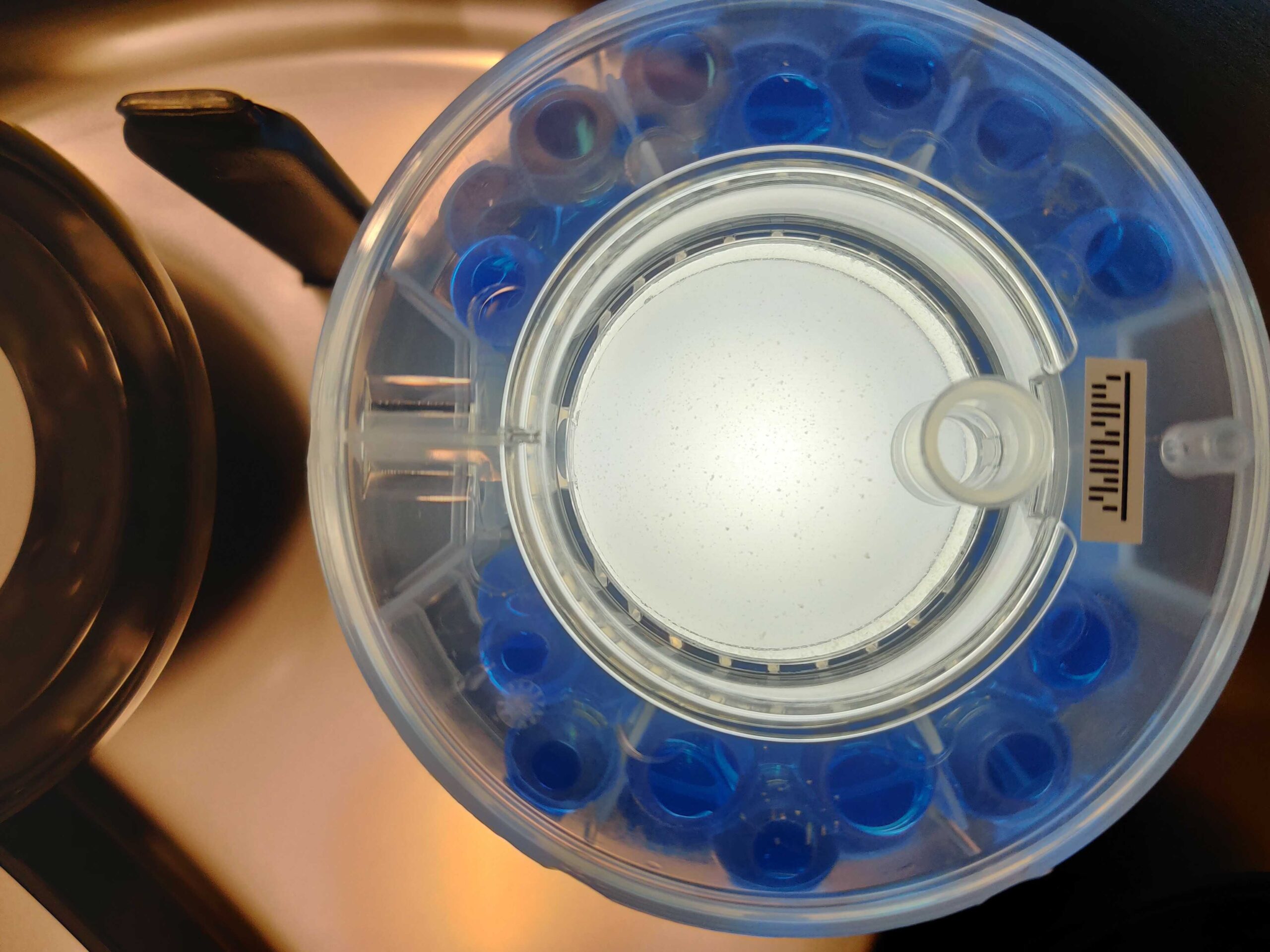
The ClinoStar’s precise control over rotational speed enables you to tailor the culture conditions. The device is versatile, allowing it to be used with different sample types, including cells (whether primary, stem cells, or cell lines), tissues, and even microorganisms.

WEBINAR
See this webinar where professor Simone Sidoli from the Albert Einstein College of Medicine discusses how he’s using 3D hepatocytes to model, treat, and analyze quiescent chromatin states.
NEWSLETTER
Keep in touch
Make sure you never miss a message.
Sign up for CelVivo’s monthly newsletter and get all the latest ClinoStar research, 3D cell culture trends, insightful symposiums, and exclusive webinars.
ClinoStar Research
Get inspired by the newest research made with the ClinoStar system on the liver.
Global Level Quantification of Histone Post-Translational Modifications in a 3D Cell Culture Model of Hepatic Tissue
DOI: 10.3791/63606
Presented below is a workflow for modeling, treating, and analyzing quiescent chromatin modifications using a three-dimensional (3D) cell culture system.
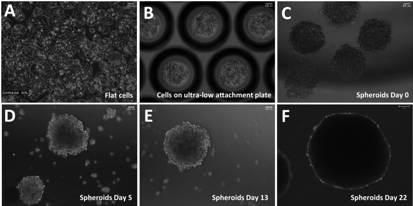
Investigation of reversible histone acetylation and dynamics in gene expression regulation using 3D liver spheroid model
DOI: 10.1186/s13072-022-00470-7
A demonstration, that 3D liver spheroids are a suitable system to model chromatin dynamics and response to epigenetics inhibitors.
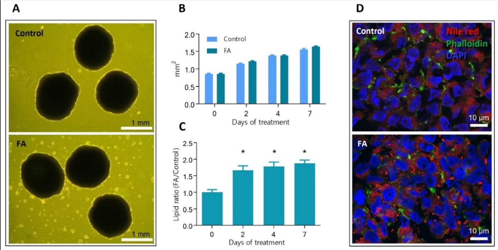
Mapping Proteome and Lipidome Changes in Early-Onset Non-Alcoholic Fatty Liver Disease Using Hepatic 3D Spheroids
We have exposed human hepatic HepG2/C3A cells-based spheroids to 65 μM oleic acid and 45 μM palmitic acid and employed proteomics and lipidomics analysis to investigate their effect on hepatocytes.
Start optimising your
In Vitro models today
