
Expand
Blastocysts with
the clinostar
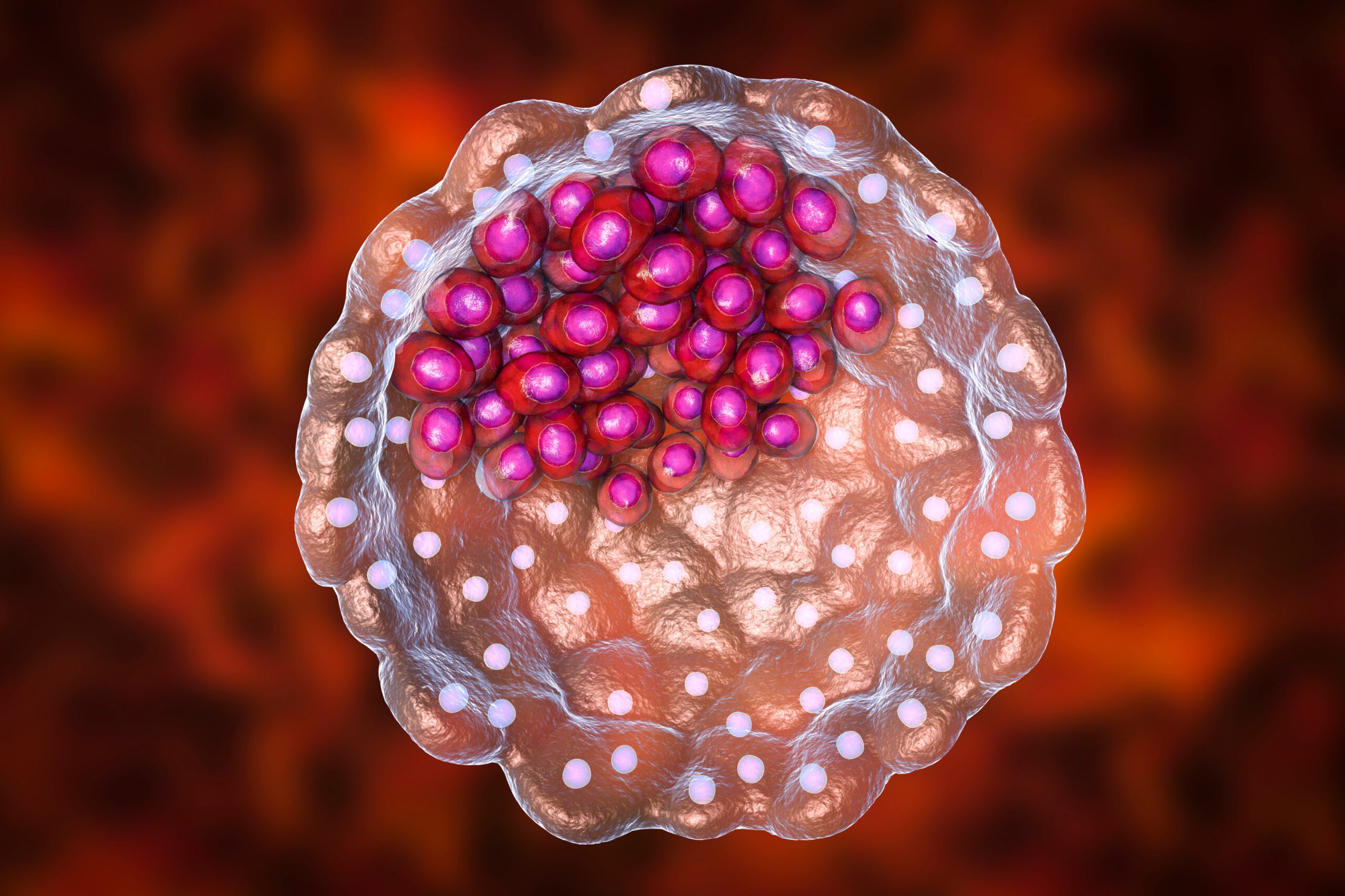
What is a blastocyst?
The blastocyst begins to form after the fertilized egg (zygote) has undergone several rounds of cell division, but before it has implanted in the uterus. It is a hollow ball of cells that forms about five days after fertilisation. The “ball” consists of an outer layer of trophoblast cells and an inner mass of cells at one side of the blastocyst that will develop into the embryo.
How to grow blastocysts with 3D cell culture
Blastoids serve as an in vitro model of the blastocyst. It is possible to culture blastoids in 3D cell culture systems for weeks in order to study their development and behavior.
There are two ways to achieve this. In one approach, a small amount of blastomere cells is isolated from a blastocyst and placed in a 3D cell culture system. The blastomeres will begin to divide and differentiate, ultimately forming a blastoid. The other approach employs pluripotent stem cells instead of blastomere cells to create the blastoid. This latter method circumvents limitations in the number of blastomere cells available.

How to work with blastocysts
in the ClinoStar
The 3D culture system provides a more physiological environment for the blastoid to grow in, as it allows for the formation of cell-cell and cell-matrix interactions. Furthermore, the environment of a 3D microenvironment is similar to the one found in the uterus.
Studying blastoids in 3D cell culture can provide insight into early embryonic development and blastomere differentiation. It can also be useful when developing strategies for improving the culture and growth of embryos in assisted reproductive technologies, such as in vitro fertilization (IVF).

ClinoStar Research
Get inspired by the newest research made with the ClinoStar system.

Bovine blastocyst like structures derived from stem cell cultures
DOI: 10.1101/2022.12.20.521301
A recent study on a newly developed and efficient method to generate bovine blastocyst-like structures (termed blastoids) via the assembly of trophoblast stem cells and expanded potential stem cells.

Investigation of reversible histone acetylation and dynamics in gene expression regulation using 3D liver spheroid model
DOI: 10.1186/s13072-022-00470-7
A demonstration, that 3D liver spheroids are a suitable system to model chromatin dynamics and response to epigenetics inhibitors.

A novel NCI-H69V small cell lung cancer functional mini-tumor model for future treatment screening applications
DOI: 10.1002/btpr.3253
In vitro cancer models are crucial in chemotherapy development, and three-dimensional (3D) models aim to bridge the gap between two-dimensional (2D) flat cultures and in vivo testing.
NEWSLETTER
Keep in touch
Make sure you never miss a message.
Sign up for CelVivo’s monthly newsletter and get all the latest ClinoStar research, 3D cell culture trends, insightful symposiums, and exclusive webinars.
Start optimising your
In Vitro models today
