Blog: Oxygen – the essential primordial stress factor
Surprisingly, the importance of oxygen is often overlooked in cell culture.
Traditionally cells are cultivated in incubators in flasks or in microtiter plates. This sets off an avalanche of hidden consequences.
Consider the journey that each molecule has to take.
The atmosphere contains about 21% oxygen. Incubators are usually humidified and contain 5% CO₂. This reduces the amount of oxygen available to 18.6% at sea level (and to 14.9% at 2,000 m above sea level) [1]. Varying humidity (as for example happens when the incubator door is opened) will lead to changes in the amount of oxygen available.
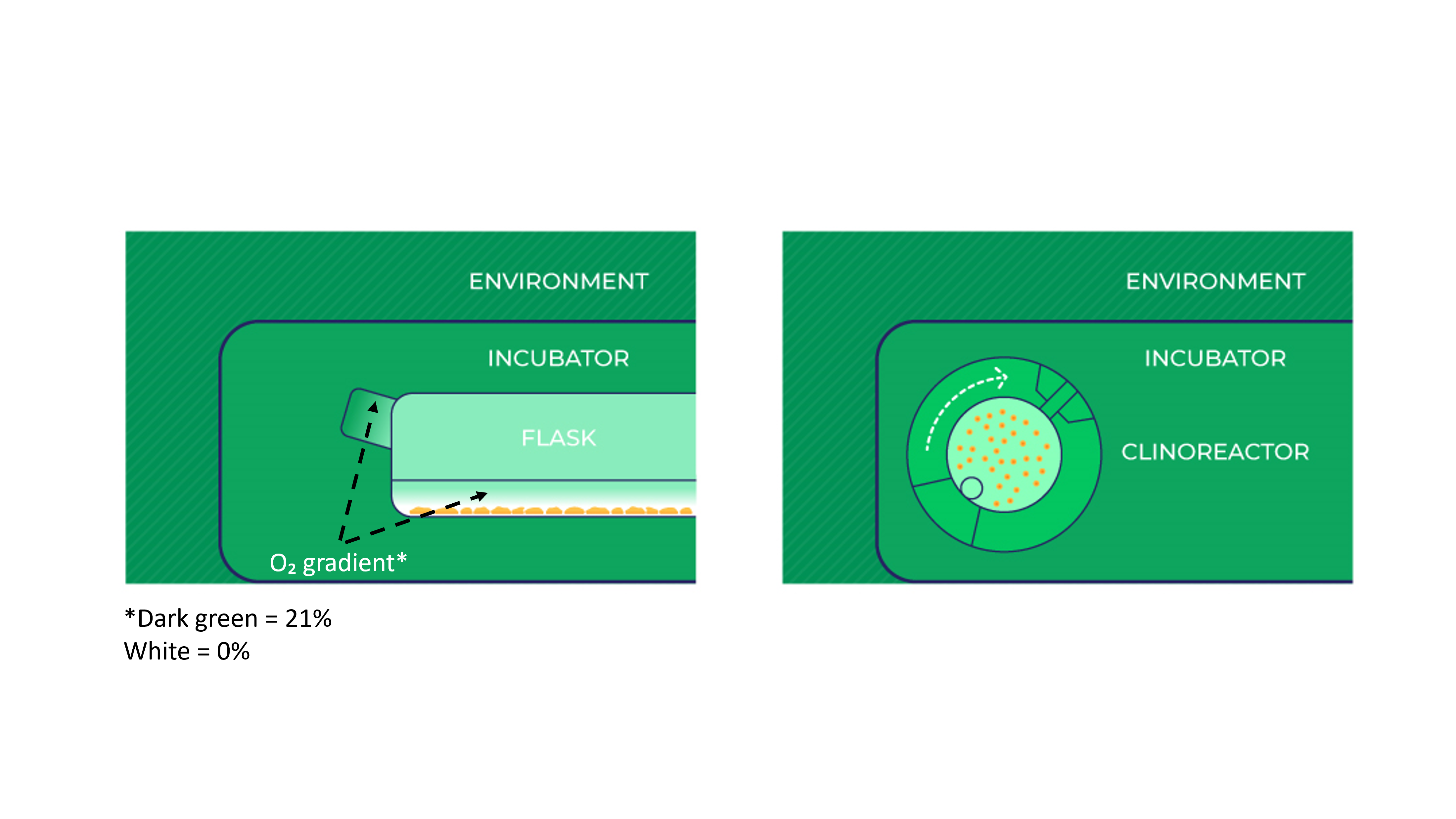
The oxygen molecule has to diffuse through a filter and into the flask or microtiter plate. Thereafter it has to penetrate the gas-media interface. The latter is strongly area dependent. Starting with an anoxic sterile medium, it can take more than 2 hrs to reach equilibrium. Bubbling air through the media (something that is rarely done) would increase the air-media contact and reduce this time to a few minutes [2] [3] [4].
Mitochondria consume oxygen. Locally, this lowers the amount of oxygen available. If the culture is vigorously growing and nearing confluency, then due to the slow rate of oxygen diffusion through media, some cells may experience a total lack of oxygen (anoxia) if the media is deeper than 3 mm. If the culture flask is not absolutely level, then some cells may have sufficient while others will suffer anoxia and initiate apoptosis! [1] [4].
Therefore it is possible that cells are being cultured in conditions where the amount of oxygen is variable and possibly scarce or even absent.
Sudden reoxygenation can cause more damage that hypoxia
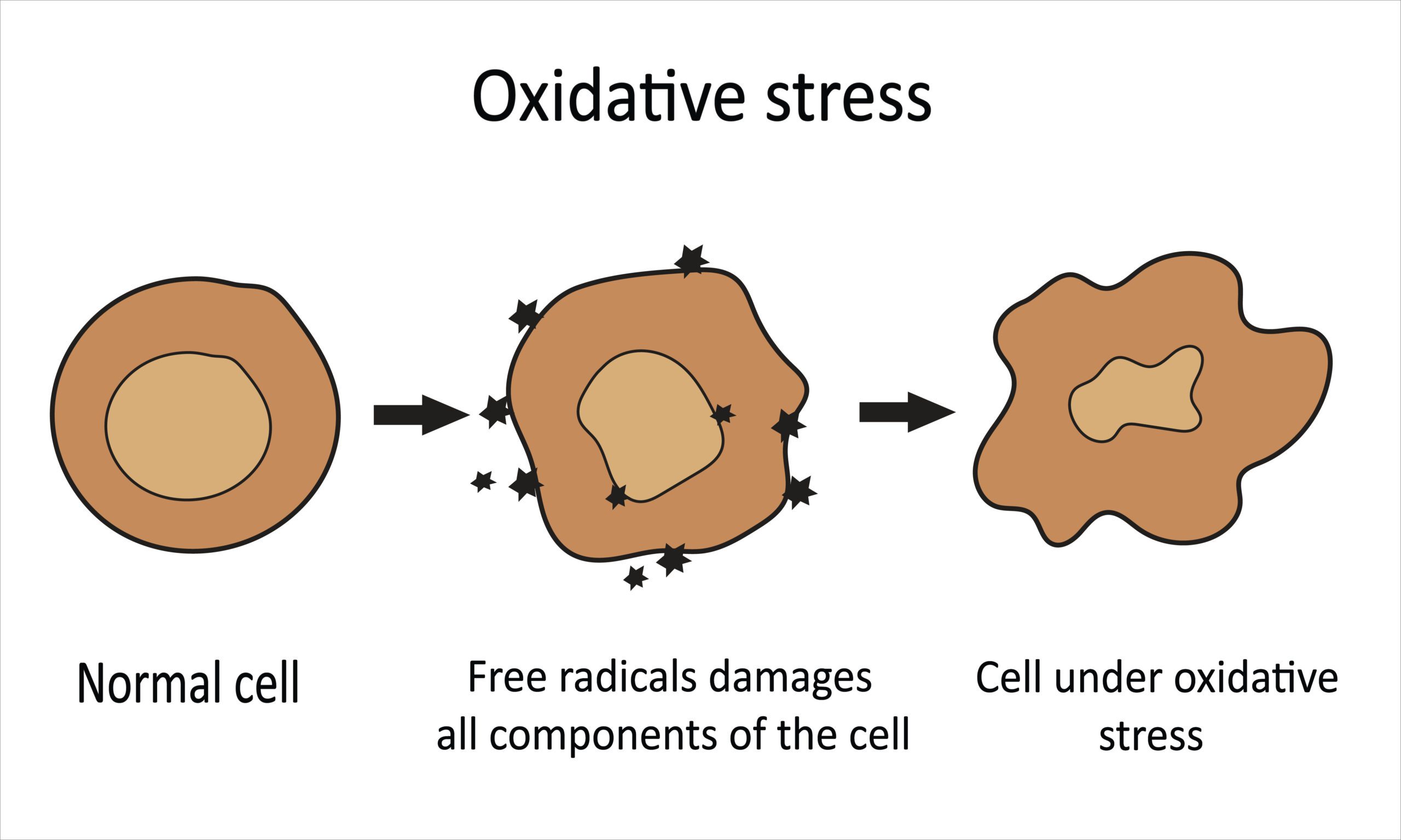
In conditions of low levels of oxygen, it is possible to suddenly re-introduce oxygen to the cells. This can occur for example when the flask is moved for any reason (e.g. inspection of cell growth, media change etc.). The movement of the media, or its renewal with fresh media, will disrupt established diffusion gradients and expose cells suddenly to ‘high’ levels of oxygen (often called an ischaemia-reperfusion event). This reoxidation can cause more damage than the hypoxia that precedes it (especially in low ATP-level or nutrient situations). It will result in the formation of high levels of reactive oxygen species (ROS, usually produced by the mitochondria), which will oxidatively damage proteins, DNA and lipids. These modifications can be reversible or irreversible and can lead to the loss of structure, function and/or enzymatic activity and increase their turnover [5] [6].
In other words oxidative stress (both the paucity of oxygen and its sudden reintroduction) can have significant harmful effects on the cell.
Get rid of diffusion gradients and oxygen depletion zones
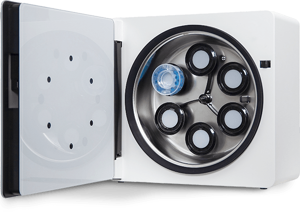
Cell culture equipment like the ClinoStar help to eliminate this oxidative stress [7].
- Firstly, rotation of the ClinoReactor induces the Coandă effect to increase the flow of air through the ClinoReactor and across a gas permeable membrane, greatly accelerating the flux of oxygen into the culture media. This maintains high levels of oxygen in the culture media at all times.
- Secondly, humidification inside the ClinoStar greatly facilitates the rapid transport of gasses (O₂ and CO₂) across the membrane. Without this humidification, the membrane would be essentially gas impermeable.
- Thirdly, the same rotation of the ClinoReactor causes the Coriolis force to gently rotate the cell clusters in the culture chamber, stirring the media and reducing the oxygen depletion zone to an absolute minimum. This effectively eliminates diffusion gradients.
- Finally, the number of times that the door needs to be opened has been minimised by the use of cameras to inspect the spheroid or organoid cultures. This reduces variations of oxygen levels in the atmosphere and in the media (and also reduces the risk of infection).
All of these ‘hidden’ features significantly increase the availability of oxygen to the cells by eliminating or reducing diffusion gradients to a minimum. This will also reduce the risk of forming reactive oxygen species (by avoiding sudden changes in oxygen availability) and thus reduce damage to DNA, proteins and lipids. Both of these features significantly improve the growth conditions.
Under these conditions the amount of oxygen available to the cell clusters is directly linked to the amount of oxygen in the ClinoStar and its flux into the culture media.

Oxidative phosphorylation or aerobic glycolysis?
In the 1920’s Warburg observed that tumours convert most of their glucose to lactate and referred to this process as aerobic glycolysis (in contrast to oxidative phosphorylation) because it occurred regardless of whether oxygen was present or absent. Since he saw the same effects in proliferating ascites tumour cells in vitro, Warburg hypothesized that metabolic reprogramming was specific to cancer cells, and was due to a mitochondrial defect. Now it is known that all tissues exhibit some degree of oxygen-independent aerobic glycolysis beside their normal oxygen-dependent oxidative phosphorylation [8].
Intracellular oxygen levels are regulated by the hypoxia inducible factor (HIF). HIF plays a key role in the cell and is known to be able to reprogram a vast array of genes involved in cellular metabolism, proliferation, extracellular matrix formation, angiogenesis and eventually can even activate mitochondrially-orchestrated apoptosis [9] [10].
The blood supply to different tissues is strikingly different (compare for example the vasculature to the brain or bone with that to spleen) [11]. A more accurate understanding of cellular physiology in different tissues requires that it is possible to reliably regulate the level of oxygen available in the media.
Controlled hypoxia in vitro
For this reason, CelVivo has introduced a hypoxia module that can regulate the amount of oxygen available in the atmosphere within the ClinoStar from ambient (i.e. probably 18-19%) to 2%. Because the diffusion gradients have been essentially eliminated (see above) this module can be thus used to directly control the oxygen available to the cell clusters.
This new module is thus a further critical step towards mimicking growth conditions within normal and cancerous tissues. A reliable and reproducible exposure of cells to oxygen levels seen in their parental tissues in vivo can thus provide a precise description of metabolic processes within a tissue and how it will respond to various types of treatment.
References
[1] T L Place, F E Domann, A J Case. Limitations of oxygen delivery to cells in culture: An underappreciated problem in basic and translational research. Free Radic Biol Med. 2017 Dec:113:311-322. doi: 10.1016/j.freeradbiomed.2017.10.003.
[2] C B Allen, B K Schneider, C W White. Limitations to oxygen diffusion and equilibration in in vitro cell exposure systems in hyperoxia and hypoxia. Am J Physiol Lung Cell Mol Physiol. 2001 Oct;281(4):L1021-7. doi: 10.1152/ajplung.2001.281.4.L1021.
[3] D Newby, L Marks, F Lyall. Dissolved oxygen concentration in culture medium: assumptions and pitfalls. Placenta. 2005 Apr;26(4):353-7. doi: 10.1016/j.placenta.2004.07.002.
[4] Z J Rogers, T Colombani, S Khan, K Bhatt, A Nukovic, G Zhou, B M Woolston, C T Taylor, D M Gilkes, Nikolai Slavov, S A Bencherif. Controlling pericellular oxygen tension in cell culture reveals distinct breast cancer responses to low oxygen tensions. bioRxiv. 2023 Oct 3:2023.10.02.560369. doi: 10.1101/2023.10.02.560369
[5] M J Sebastião, P Gomes-Alves, I Reis, B Sanchez, I Palacios, M Serra, P M Alves. Bioreactor-based 3D human myocardial ischemia/reperfusion in vitro model: a novel tool to unveil key paracrine factors upon acute myocardial infarction. Transl Res. 2020 Jan:215:57-74. doi: 10.1016/j.trsl.2019.09.001
[6] J A Trujillo-Hernandez, R L Levine. Response to oxidative stress of AML12 hepatocyte cells with knockout of methionine sulfoxide reductases. Free Radic Biol Med. 2023 Aug 20:205:100-106. doi: 10.1016/j.freeradbiomed.2023.05.028.
[7] K Wrzesinski, S J Fey. Metabolic Reprogramming and the Recovery of Physiological Functionality in 3D Cultures in Micro-Bioreactors. Bioengineering (2018), 5(1), 22; doi:10.3390/bioengineering5010022
[8] M Jaworska, J Szczudło, A Pietrzyk, J Shah, S E Trojan, B Ostrowska, K A Kocemba-Pilarczyk. The Warburg effect: a score for many instruments in the concert of cancer and cancer niche cells. Pharmacol Rep. 2023 Aug;75(4):876-890. doi: 10.1007/s43440-023-00504-1.
[9] K E R Hollinshead, D A Tennant. Mitochondrial metabolic remodeling in response to genetic and environmental perturbations. Wiley Interdiscip Rev Syst Biol Med. 2016 Jul;8(4):272-85. doi: 10.1002/wsbm.1334.
[10] A Yfantis, I Mylonis, G Chachami, M Nikolaidis, G D Amoutzias, E Paraskeva, G Simos. Transcriptional Response to Hypoxia: The Role of HIF-1-Associated Co-Regulators. Cells. 2023 Mar 3;12(5):798. doi: 10.3390/cells12050798.
[11] H M Swartz, A B Flood, P E Schaner, H Halpern, B B Williams, B W Pogue, B Gallez, P Vaupel. How best to interpret measures of levels of oxygen in tissues to make them effective clinical tools for care of patients with cancer and other oxygen-dependent pathologies. Physiol Rep. 2020 Aug;8(15):e14541. doi: 10.14814/phy2.14541
In vivo just moved closer
Learn more about how you can benefit from using the ClinoStar and clinostat principle when culturing cells in your lab.


