Spheroid Characteristics

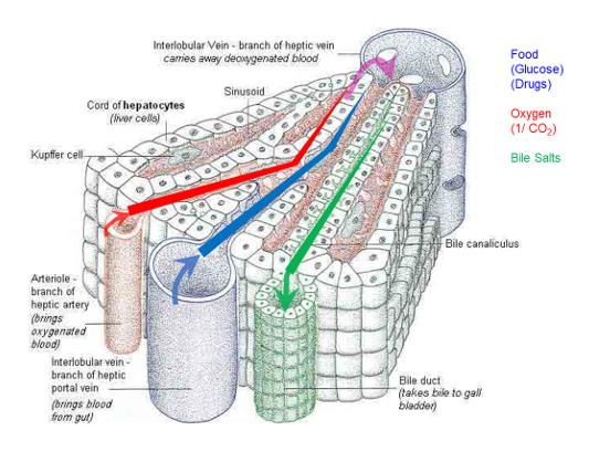
The individual cells in vivo will experience different exposure to nutrients, oxygen, CO2 oxygen and bile salts. Below is displayed a drawing of these characteristics for a liver.
Spheroids formed in the CelVivo system will after approximately 3 weeks act like cells in vivo and have developed ultrastructure which resembles tissue. The cells will have the same characteristics as the cells in vivo.
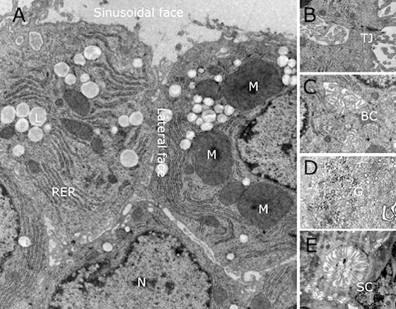
The spheroids have not only gained functionality of in vivo conditions but also developed architecture and structure as such. This is exemplified in this electron microscope picture.
In order for any model to be truly beneficial the model needs to be reproducible. Spheroids grown in our system has s very high reproducibility. The fact that the high reproducibility together with unmatched correlation with in vivo behavior of spheroids grown in our system makes them the ideal models of tissue.

Lipid droplets, M Mitochondria, RER Rough endoplasmic, reticulum, N Nucleus, TJ tight junctions
BC Bile canaliculus-like channel, G Glycogen granules, SC Sinusoidal-like channel
| SPHEROIDS | Characteristics For HepG2-C3A – values for other cell lines will be different |
| Number of cells per spheroid (± 22%) |
|
| Spheroid size (Planimetric surface) |
|
| Protein concentration |
|
| ATP concentration |
|
| Bioreactor soluble protein content |
|
